Crystal Voilet (Gram’s) solution for microscopy
Description
Crystal Violet (Gram's) solution for microscopy is a purple aqueous primary stain (typically 1% crystal violet in water or alcohol) used in the Gram staining technique to differentiate Gram-positive (purple) from Gram-negative (pink/red) bacteria based on cell wall peptidoglycan differences.
Composition and Preparation
Primary component: Crystal violet dye (gentian violet, CI 42555), often with ammonium oxalate for stability.
Standard formulation: 1% crystal violet solution, clear purple liquid suitable for microscopic staining.
Role: Initial stain binds to all bacterial cells; fixed by Gram's iodine mordant.
Staining Procedure
Prepare thin heat-fixed bacterial smear on slide.
Flood with Crystal Violet (Gram's) for 30–60 seconds, rinse gently with water.
Apply Gram's iodine 30–60 seconds, rinse; decolorize with ethanol/acetone 10–20 seconds, rinse; counterstain with safranin 30–60 seconds, rinse, dry, observe under oil immersion.
Results: Gram-positive retain violet (thick peptidoglycan); Gram-negative lose dye, take safranin (pink).
Properties and Handling
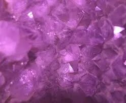
Appearance: Purple solution, light-sensitive; store dark.
Critical factor: Decolorization timing affects results; over-decolorizes Gram-positive to pink.
For catalog: “Crystal Violet (Gram’s) solution for microscopy, 1% purple stain, for bacterial differentiation in Gram procedure; Gram+ purple, Gram- pink/red
Specifications
| Model No. | N-CH1651 |
|---|